Ultrasound tech’s discovery paved way for swift treatment
“Immediately, something didn’t really look right”
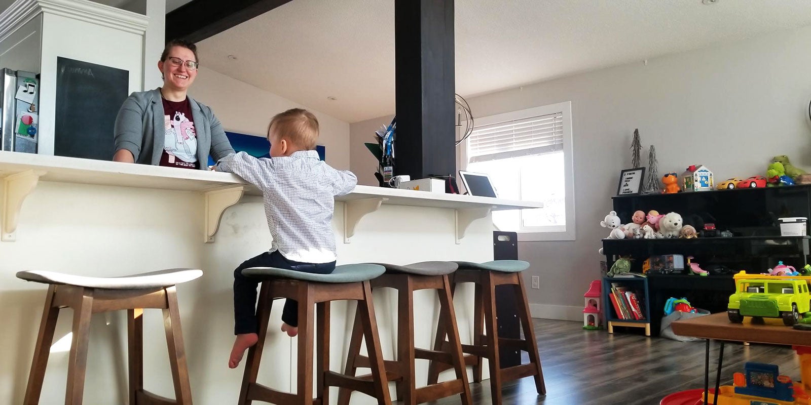
Two-year-old Artie grins and hikes up his shirt, revealing the pale vertical scar on his chest. To him, it’s something that’s always been there – a birthmark of sorts.
For his parents, though, it’s a reminder of the complex heart surgery that saved his life.
Artie’s show-and-tell moment passes quickly, and – as if to show he’s no different from most toddlers – he roots around his collection of toy cars in the family’s living room.
“Po-lice car,” he offers. “Will you play with it?”
Artie’s life-saving operation, just days after his birth, was the culmination of a chain of events that started months before, with an obstetric, or prenatal, ultrasound in May of 2020.
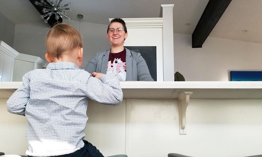
“I wasn’t expecting anything to be wrong with him,” says his mother, Amy Porter. “That happens to other people. And then suddenly, it was me.”
In fact, it’s a diagnosis that will affect a relatively large number of families. Congenital heart disease affects about 1 in 100 babies born in Canada, making it the most common birth defect. In Alberta, more than 40% of major heart conditions are not detected before birth, with the problem more pronounced in rural areas.
That’s why Porter is now working to raise awareness of the need to improve detection rates through prenatal ultrasounds – highlighting an important and often overlooked aspect of health care. It’s a mission that began, in part, because of what a NAIT-educated ultrasound technologist did for her son.
“It’s not [just about] finding out what flavour of baby you’re going to have,” Porter says. “They’re checking up on your child’s well-being.”
The day everything changed
Early on, Porter’s pregnancy had been uneventful, and she was told her previous ultrasounds were normal. It was a relief after earlier attempts to get pregnant using donor sperm had failed.
Then, at 28 weeks gestation, Porter’s obstetrician told her that her belly was measuring larger than expected and sent her for a follow-up scan.
That ultrasound changed everything.
Covid restrictions meant Porter’s wife, Sam, and their three-year-old daughter couldn’t join her in the exam room with ultrasound technologist Whitney Lane (Diagnostic Medical Sonography ’08).
“Because it was only me, I was a little less chatty,” says Porter. Part way through the scan, she felt prickles of unease. “We were looking at his heart. And [the technologist] spent a long time looking at it.”
Later, alone in the room while Lane filed her report, Porter’s anxiety swelled.
“I was like, something’s wrong with his heart. I knew … something’s not right. This isn’t good.”
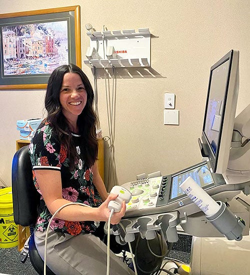
Down the hall, Lane discussed her findings with the clinic’s radiologist. The exam, which checks on the growth and well-being of babies in the third trimester, is one she has scanned hundreds of times over her 14-year career at MIC Medical Imaging, a diagnostic imaging provider in the greater Edmonton area.
“Immediately, something didn’t really look right,” Lane says.
When scanning the heart, sonographers check for several features, including how the large vessels that pump blood to the lungs and body are situated.
“He was in such a good position that it should have been easy,” she says. “But I couldn’t make [it] look normal.”
Lane believes working as a temporary contractor in New Zealand – she’s currently on her sixth stint there – helped her detect Artie’s heart anomaly with confidence. Guidelines for obstetrical exams are more stringent in the island country, she notes.
“I do have a little bit more experience,” she says, “because when I’m here [in New Zealand] I have to do such an extensive view of the heart.”
The next day, Porter’s obstetrician told her the baby had a rare heart condition, and handed her a yellow sticky note with the words “Transposition of the Great Arteries” or TGA – a fatal condition if untreated.
Artie’s specific condition, d-TGA – in which the major vessels of the heart are reversed – is rare, accounting for 5% to 7% of all congenital heart defects. This disrupts the normal flow of blood, depriving the body of oxygen.
Initially dismayed, Porter says she and her wife were relieved to learn that surgical intervention in the first few days of life has a 96% success rate. But had Artie’s condition not been detected by Lane during that prenatal ultrasound, Porter is convinced he would not have survived.
“He wouldn’t be here, I am certain,” she says. “When he was born, he was blue … he was greyish. He didn’t cry, he was totally quiet. Two-and-a-half hours after he was born, he crashed.”
At the Stollery Children’s Hospital, surgeons urgently created a passage between the chambers of Artie’s heart so that some oxygenated blood could reach the rest of his body.
Six days later, he underwent open-heart surgery.
On a mission

Today, Porter has turned her son’s diagnosis into a mission to help other families.
With support from fetal and pediatric cardiologists Dr. Lisa Hornberger and Dr. Luke Eckersley, she established a non-profit foundation called Tiny HeartsCan last fall. She aims to improve prenatal detection rates by raising awareness, as well as funding to provide enhanced training for sonographers.
Hornberger, a renowned leader and researcher in fetal and pediatric echocardiography, or heart ultrasounds, attests to the importance of prenatal detection. She knows the impact it has on successful outcomes.
“Knowing ahead of time means the families can be prepared, the moms can be prepared, the babies are delivered in the right place, and the right things are done for the baby from the beginning, so they stay nice and stable and healthy before going to surgery,” says the divisional director of cardiology at The Stollery.
Porter points out that expectant parents also need to recognize the significance of ultrasounds, particularly the anatomy scan at 18 to 20 weeks, when organs and systems have developed enough that they can be visualized and examined. Porter acknowledges that the way she approached that scan may have contributed to Artie’s initial missed diagnosis.
“I chatted through the whole thing like it was a hair appointment,” she recalls in disbelief.
It’s a balancing act for sonographers, says Martie Grant, obstetrics lead for NAIT’s Diagnostic Medical Sonography program. Students are taught to be clear with patients about the need to focus on the exam itself, she explains, before the more casual, show-and-tell aspect.
As Porter’s case demonstrates, the work of sonographers does affect someone’s life, she says.
“Everything that they do has an implication on how that pregnancy is managed, what happens next for the patient, what happens next for the baby,” Grant says.
At his family’s home, as Artie jogs around the kitchen, bare feet slapping the floor, there’s no evidence of his traumatic beginning. “Chase me, Mommy!” he squeals.
He’s a healthy, energetic child, but his mother points out his journey is far from over. Cardiologists have detected some narrowing to one of his pulmonary artery branches due to scar tissue, and Porter worries he may eventually have to undergo another open-heart surgery.
Despite the uncertainty over Artie’s health, she says she’s grateful that Lane’s diligence has led her down a path to helping others.
“Being a parent was what I was meant to do and [Artie’s] diagnosis has given me purpose,” Porter says.
A key role in health care

Artie’s journey has been meaningful for sonographer Lane, as well. Throughout her career, she estimates she’s detected five major heart defects, but says Artie’s case is the one that stands out. Months after his birth, Lane received a handwritten thank-you letter from Porter.
That acknowledgement reaffirmed that her profession plays a key role in the health-care system.
“We do have the power to catch things that could potentially save a life,” Lane says. “A lot of the time, you don’t really know if you’re making a difference, and so I think it … kind of re-energized my career and made me feel grateful for what I do and that I can do it.”
Reaching out to Lane was important, Porter says, because her work paved the way for her son’s successful outcome and, through the foundation, that of other infants in the future. “No words will ever be enough.”
Northern Alberta Institute of Technology
Mailing Address:
11762 - 106 Street
Edmonton, AB, Canada, T5G 2R1
At NAIT, we honour and acknowledge that the land on which we learn, work and live is Treaty Six territory. We seek to learn from history and the lessons that have come before us, and to draw on the wisdom of the First Peoples in Canada. Only through learning can we move forward in truth and reconciliation, and to a better future together. Read more
About techlifetoday
Techlifetoday is the source for NAIT alumni and students to discover news about the polytechnic, and insights, advice and inspiration from the NAIT community to help them build successful careers and lead fulfilling lives.